Prashanta Dutta, assistant professor in the School of Mechanical and Materials Engineering, is trying to thread the eye of a tiny needle. But, instead of the 1,230 microns of an average-sized needle eye, Dutta’s is only 10 microns wide, and the “thread’’ is five microns wide.
Dutta and his colleagues recently received a grant from the National Institute of Health’s National Institute for Biomedical Imaging and Bioengineering for the development of a novel biosensor that would identify biological materials more quickly than current methods. The research could have applications for human health, environmental monitoring and homeland security.
During the past several years, researchers have sequenced human DNA through the human genome project. To better understand human biology, however, requires understanding of the tiny proteins that provide functionality for the DNA.
“The structure of the protein determines how you behave,’’ says Dutta. Separating and identifying proteins will help researchers better understand their function, providing benefit to the biochemical and medical fields.
For the past 50 years, researchers have used two methods to separate proteins, electrophoresis and mass spectrometry. A mass spectrometer separates materials by atomic mass, but the machine is the size of a large cabinet. Electrophoresis effectively separates proteins using an electric field, but the process is slow.
So, for instance, during the SARS (severe acute respiratory syndrome) scare of 2003, airline passengers with a cold who came from China were quarantined for several days while their blood was tested for the SARS virus.
For the past two years, Dutta has been working to develop a device that separates tiny proteins as small as a micrometer from each other. He hopes that this hand-held device will someday do such testing in five to 10 minutes — less than the time it takes airplane passengers to go through customs.
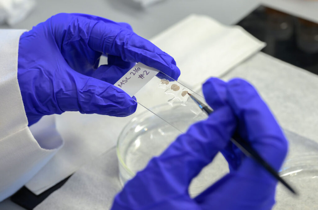
Dutta uses a novel technique that puts the proteins into a liquid, then pulls them through tiny micro-channels. Reducing the size of the separation process allows the researchers to do separations in five to 10 minutes versus several days.
They are building the tiny channels on a substrate made of poly-di-methyl siloxane (PDMS). The material is better than other silicon based materials such as glass, acrylic or silicon wafer because it can withstand high voltage without leaking current. However, it bends easily, which means that the nanosized electrodes deposited on tiny channels break when the material is inadvertently flexed.
“And everyone wants to bend it — from the university professor on down to kids,’’ says Dutta.
The researchers recently have solved the problem by adding a hard silicon base to the bottom of the PDMS layer.
One of the major remaining challenges of working with soft PDMS is the precise positioning of nanoelectrodes in the microchannel. Recently this group developed a recipe to form platinum nanoelectrodes on selected locations of the microdevice using lift off-sputtering technique.
Similar to the idea of stenciling, this technique involves placing a mask over the surface of the PDMS and then using ultraviolet light to etch the desired patterns on the material.
Next Story
WSU Global Campus student ambassador shines during #CougsGive Day
Aunjelique Andersen, a fully online student pursuing her degree in Media Innovation, ranked first on the #CougsGive Ambassador Leaderboard, inspiring 54 gifts through her outreach.
Recent News
WSU SURCA undergraduate researchers earn 65 awards
Seventy‑three students who participated in the Showcase for Undergraduate Research and Creative Activities 2026 were awarded prizes totaling $14,000.
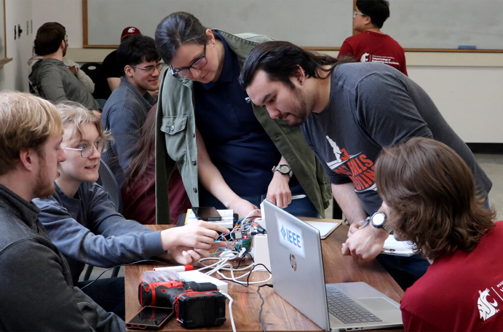
Inside WSU’s student-run hackathons
Hackathons have become a defining space for student innovation, with two taking center stage this year.
Undergrad researchers drive discovery in the College of Veterinary Medicine
Hands‑on research opportunities in the college are enabling students to contribute directly to major discoveries, including a recent Nature study.
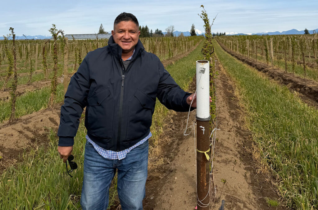
New WSU raspberry breeder to continue career goal of helping farmers
Germán Sandoya-Miranda aims to develop improved varieties and work closely with growers to help Washington farmers boost yields and adapt to evolving challenges.
WSU Regent, longtime civic leader Sam Hunt dies following battle with leukemia
Hunt, a 1967 WSU graduate, devoted more than five decades to public service, education, and civic leadership across Washington.
WSU recognized for support of first-generation students
The university’s elevation to FirstGen Forward Network Champion reflects growing enrollment, improved retention, and expanded support programs helping first-generation students succeed.